Pupillometry, the measurement of pupil size and response to light, has become an essential diagnostic tool in ophthalmology, neurology, and general medicine. Once limited to subjective penlight exams, pupil testing has advanced into a data-driven discipline supported by digital imaging, infrared sensors, and AI-powered analysis.
Modern pupillometry provides objective insight into visual pathways, neurological function, and autonomic nervous system health. As digital diagnostics evolve, the pupil now serves as a measurable indicator of both ocular and brain function rather than a simple reflex response.
1. The Science Behind Pupillometry
Pupillometry evaluates how the pupil reacts to changes in light intensity, offering insight into both afferent and efferent visual pathways.
Key metrics measured in modern pupillometry include:
- Baseline pupil diameter measured in millimeters
- Constriction latency indicating response initiation time
- Constriction velocity measuring speed of pupil contraction
- Dilation velocity reflecting recovery after light removal
- Anisocoria detection identifying inter-eye pupil size differences
Quantitative measurements replace subjective observation with repeatable clinical data.
2. From Manual Exams to Automated Measurement
Traditional pupil assessments relied heavily on clinician judgment, resulting in variability between examiners. Automated pupillometry replaces estimation with precision by capturing real-time pupil dynamics digitally.
Advantages of automated pupillometry include:
- Standardized testing that reduces examiner bias
- Millisecond-level detection of pupil response changes
- Detailed waveform and trend analysis
- Digital storage for longitudinal patient monitoring
Automated systems elevate pupillometry from observation to measurable clinical evidence.
3. The Growing Role of AI in Pupillometry
Artificial intelligence now plays a central role in pupil analysis by detecting subtle abnormalities that may not be visually apparent.
AI-enhanced pupillometry supports:
- Identification of afferent pupillary defects
- Early detection of neurological or optic nerve dysfunction
- Pattern recognition across large datasets
- Predictive modeling of disease progression
AI strengthens diagnostic accuracy while supporting clinician decision-making.
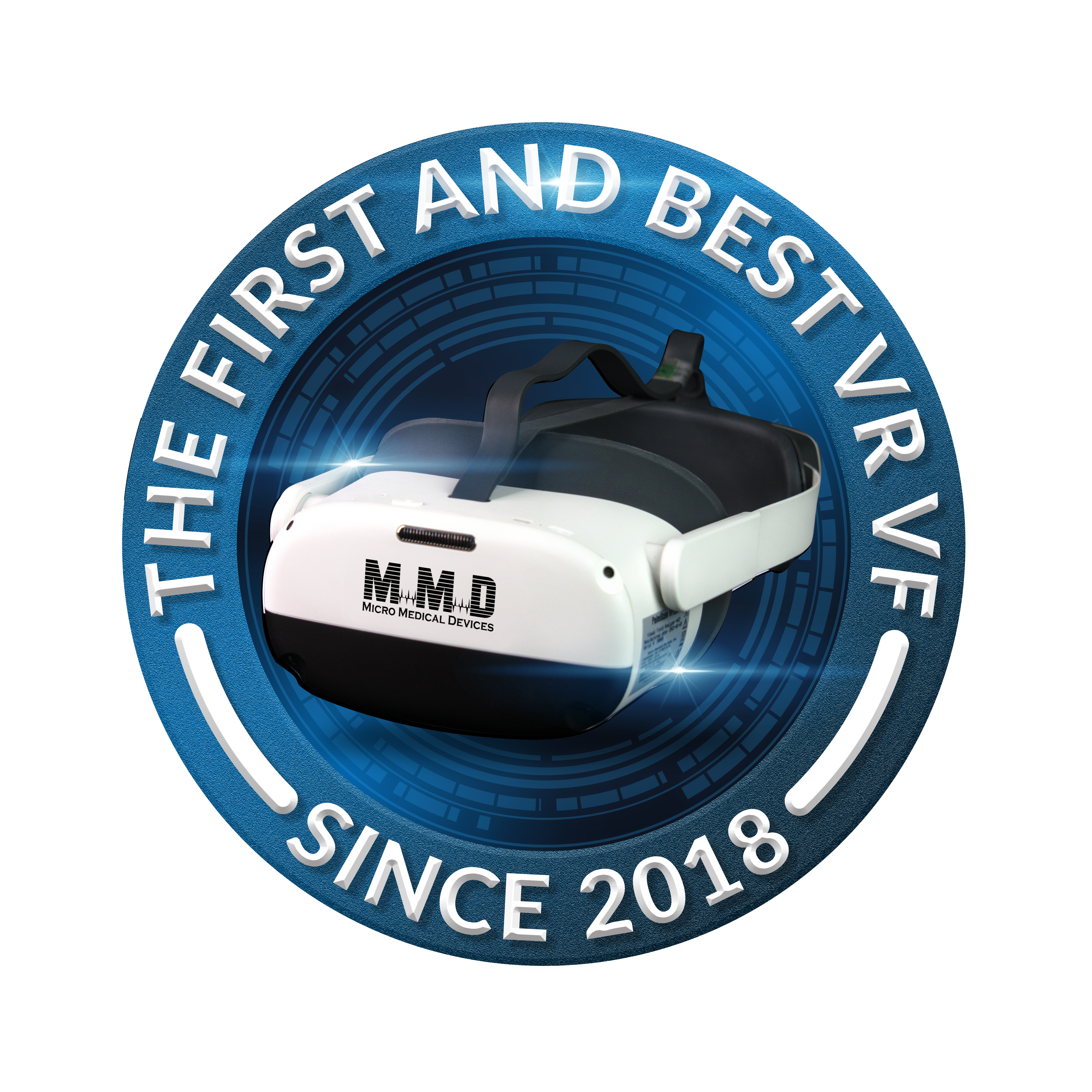
4. Integration with Virtual Reality Visual Field Testing
Innovative platforms are exploring the integration of pupillometry into virtual reality visual field testing environments. By combining pupil response tracking with functional vision assessment, clinicians gain deeper insight during a single exam.
Benefits of integrating pupillometry with VR perimetry include:
- Simultaneous evaluation of pupil response and visual field sensitivity
- Improved detection of visual pathway abnormalities
- Non-invasive and comfortable headset-based testing
- Portable diagnostics suitable for clinic or remote use
Virtual Field is among the platforms advancing this combined functional and physiological testing approach.
5. Clinical Applications of Modern Pupillometry
Pupillometry is now widely used across multiple clinical disciplines to support diagnosis and monitoring.
Common clinical applications include:
- Glaucoma assessment through detection of afferent pupillary defects
- Optic neuritis evaluation and progression monitoring
- Concussion and traumatic brain injury screening
- Measurement of pharmacologic dilation response
- Autonomic nervous system imbalance assessment
When combined with visual field testing and imaging, pupillometry enhances diagnostic confidence.
6. The Impact of Portable Pupillometry Devices
Handheld and portable pupillometers have significantly expanded access to quantitative pupil testing.
Key advantages of portable pupillometry include:
- Rapid bedside assessment in hospitals and emergency settings
- Objective neurological screening in remote or mobile clinics
- Seamless cloud integration for record sharing
- Improved accessibility for pediatric and mobility-limited patients
Portability allows pupillometry to extend beyond traditional exam rooms.
7. Coding and Documentation for Quantitative Pupillometry
Quantitative pupillometry is recognized for reimbursement under CPT code 95919 when properly documented.
Required documentation elements include:
- Patient symptoms and clinical indication
- Quantitative measurements such as latency and velocity
- Comparison to established normative data
- Clinical interpretation and provider signature
Accurate documentation ensures both compliance and reimbursement sustainability.
8. Complementing Structural and Functional Diagnostics
Pupillometry delivers the greatest value when combined with diagnostic tools that assess ocular structure and visual function.
Common complementary technologies include:
- Virtual Field VR perimetry for functional vision assessment
- Biometry and A-Scan to evaluate ocular structure
- Pachymetry to support glaucoma risk evaluation
- Keratometry to assess corneal optics affecting light entry
- B-Scan imaging to rule out posterior segment pathology
- CXL assessment when corneal stability is clinically relevant
Diagnostic platforms from providers such as Micro Medical Devices support accurate structural measurements that strengthen pupillometric interpretation.
9. What the Future Holds for Pupillometry
The future of pupillometry lies in deeper AI integration, telehealth expansion, and multi-modal diagnostic platforms.
Emerging developments include:
- AI-based risk prediction for optic neuropathy
- Combined visual field and pupillometry testing workflows
- Cloud-based longitudinal pupil response tracking
- Integration with wearable and headset-based diagnostics
Pupillometry is evolving from measurement to meaningful clinical insight.
10. Conclusion
Modern pupillometry has transformed pupil evaluation into a precise, data-driven diagnostic discipline. Through digital automation, AI analytics, and integration with virtual reality visual field testing, clinicians can better understand the eye-brain connection and improve patient outcomes.
When combined with complementary tools such as VR perimetry, biometry, Pachymetry, Keratometer, B-Scan, and CXL, pupillometry becomes a critical component of comprehensive vision and neurological assessment.
Enhance your diagnostic workflow with modern pupillometry and advanced vision testing solutions.
Call us today to learn more about integrating data-driven pupil assessment into your practice.