Ptosis, commonly known as drooping of the upper eyelid, is not just a cosmetic issue. When the eyelid descends far enough, it can block the upper portion of a patient’s visual field and interfere with daily activities such as driving, reading, and navigating stairs.
For clinicians, accurate diagnosis and objective documentation are critical. This is especially important when determining medical necessity for surgical correction. The Superior 36 visual field test plays a key role by precisely measuring superior visual field loss and translating patient complaints into quantifiable clinical data.
The Superior 36 test provides clear, reproducible evidence of functional vision impairment and supports confident clinical decision-making.
1. Understanding Ptosis
Ptosis occurs when the upper eyelid margin sits lower than normal and partially or fully covers the pupil. It may affect one or both eyes and can develop for several reasons.
Common forms of ptosis include:
- Aponeurotic ptosis caused by age-related stretching of the levator muscle
- Neurogenic ptosis associated with nerve conditions such as oculomotor nerve palsy or Horner syndrome
- Myogenic ptosis is related to muscle disorders like myasthenia gravis
- Mechanical ptosis caused by eyelid tumors, swelling, or excess tissue
- Congenital ptosis is present at birth due to abnormal levator development
Identifying the underlying cause is important, but measuring how ptosis affects vision is what completes the diagnosis.
2. Why Visual Field Testing Is Critical in Ptosis
Ptosis reduces functional vision by blocking the superior visual field. This loss must be objectively measured to determine clinical significance and eligibility for surgical intervention.
The Superior 36 visual field test focuses on the area most affected by eyelid droop.
Key reasons visual field testing matters in ptosis include:
- Objective measurement of superior visual field loss
- Documentation of functional impairment for medical records
- Support for surgical justification and insurance approval
- Baseline data for postoperative comparison
Objective visual field results transform subjective symptoms into defensible clinical evidence.
3. What Is the Superior 36 Visual Field Test
The Superior 36 test is a specialized visual field pattern designed to evaluate the upper portion of the visual field using 36 targeted test points.
This test concentrates on the superior field where ptosis has the greatest impact.
Key characteristics of the Superior 36 test include:
- Focus on the upper third of the visual field
- Sensitivity to mild and moderate superior field obstruction
- Compatibility with threshold and suprathreshold testing
- Reliable comparison for preoperative and postoperative assessment
Unlike standard visual field patterns, the Superior 36 isolates the functional loss that matters most in ptosis evaluation.
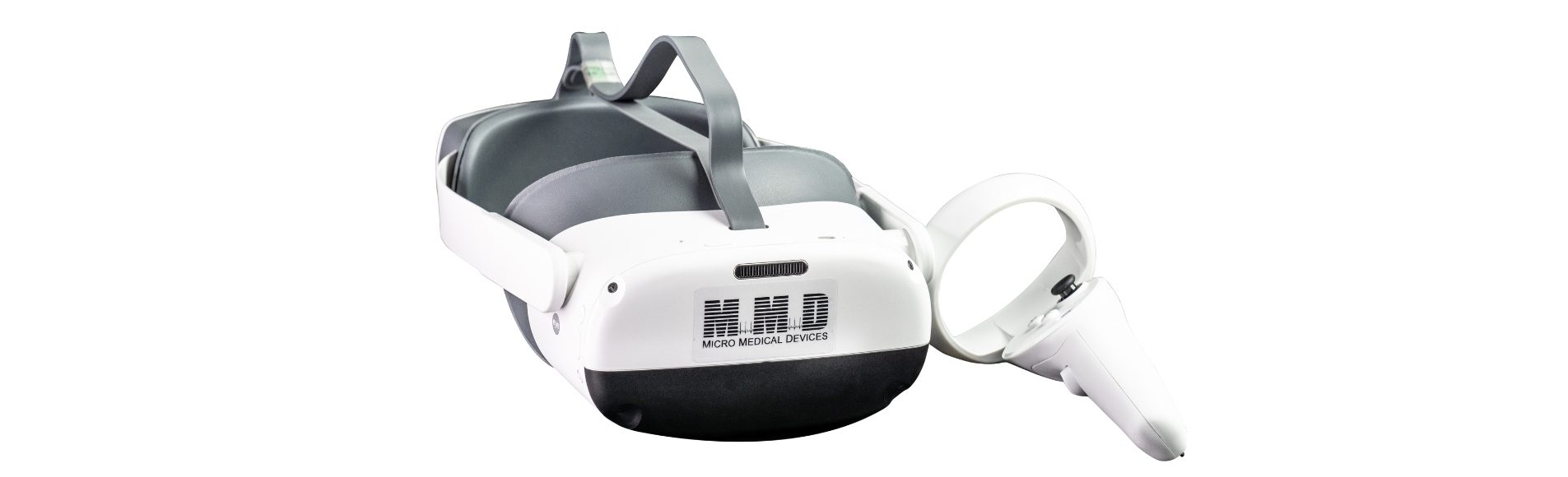
4. The VF2000 Perimetry in Ptosis Evaluation
Traditional perimetry systems can be uncomfortable and restrictive for patients with eyelid droop. VR perimetry offers a more flexible and patient-friendly alternative.
The VF2000 enables accurate Superior 36 testing using a portable headset-based system.
Advantages of using The VF2000 for ptosis assessment include:
- Dedicated Superior 36 testing mode
- AI-assisted fixation monitoring for improved reliability
- Cloud-based reports are suitable for documentation and insurance submission
- Comfortable testing posture that reduces eyelid fatigue
- Easy comparison of preoperative and postoperative results
Virtual perimetry allows clinicians to perform advanced ptosis testing without the limitations of traditional equipment.
5. Clinical Workflow for the Superior 36 Test
Incorporating the Superior 36 test into clinical practice is straightforward when using modern visual field platforms.
Typical workflow steps include:
- Preparing the patient with clear fixation instructions
- Selecting the Superior 36 protocol in the testing software
- Performing baseline testing before surgical planning
- Saving and attaching results to the patient record
- Repeating the same test pattern after surgery for comparison
Consistency in testing ensures reliable clinical and administrative outcomes.
6. Medical Necessity and Insurance Documentation
Insurance providers often require objective proof of functional vision loss before approving ptosis or blepharoplasty surgery.
The Superior 36 visual field test is a cornerstone of this documentation.
Required elements often include:
- Visual field results showing significant superior field loss
- Clinical photographs demonstrating eyelid position
- Documented functional complaints such as impaired driving vision
- Postoperative visual field improvement using the same test pattern
Clear documentation protects both the patient and the practice.
7. Supporting Diagnostics in Ptosis Evaluation
While the Superior 36 test measures functional loss, additional diagnostics help rule out contributing factors and support comprehensive care.
Common complementary diagnostic tools include:
- Biometry and A-Scan measurements to assess ocular dimensions and rule out pseudo-ptosis
- Pachymetry to evaluate corneal thickness before surgical planning
- Keratometry to monitor corneal curvature changes related to eyelid weight
- B-Scan ultrasound to exclude orbital or posterior causes of lid droop
- CXL assessment when corneal stability is a concern
Diagnostic instruments from manufacturers such as Micro Medical Devices support accurate measurement and reliable clinical evaluation.
8. Advantages of VR Visual Field Testing for Ptosis Patients
VR visual field testing offers meaningful benefits for patients undergoing ptosis evaluation.
Key advantages include:
- Natural testing posture that improves comfort
- Portable setup suitable for small clinics and outreach settings
- Multilingual audio guidance for improved patient understanding
- Cloud storage for secure and accessible documentation
- Consistent calibration for repeatable results
Patient comfort directly contributes to test accuracy and reliability.
9. Postoperative Assessment Using Superior 36
Following ptosis repair or blepharoplasty, repeating the Superior 36 test provides objective confirmation of surgical success.
Postoperative assessment typically includes:
- Performing the same Superior 36 protocol used preoperatively
- Comparing the percentage of superior field restoration
- Documenting improvement in functional vision metrics
- Storing results in the patient record for audit readiness
The Superior 36 test delivers clear before-and-after evidence of functional improvement.
10. Conclusion
Accurate evaluation of ptosis requires more than visual inspection. Functional vision loss must be objectively measured, documented, and tracked.
The Superior 36 visual field test remains a gold standard for assessing superior visual field obstruction. When delivered through The VF2000 VR perimetry, clinicians gain a precise, patient-friendly, and efficient diagnostic solution.
Combined with complementary diagnostics such as biometry, Pachymetry, Keratometer, B-Scan, and CXL, the Superior 36 test supports a complete and evidence-based approach to ptosis diagnosis and management.
Improve ptosis diagnosis and documentation with precise superior visual field testing.
Call us today to learn how The VF2000 and advanced diagnostic tools can support accurate assessment, surgical planning, and insurance compliance.