Assessing the visual field is an essential part of every comprehensive eye examination. The Confrontation Visual Field Test remains a quick and cost-effective method to detect gross visual field defects, such as scotomas or hemianopias, often associated with optic nerve or brain lesions. While newer technologies like VR visual field and virtual perimetry are transforming how clinicians assess vision, the confrontation method remains a valuable frontline screening tool — especially when digital tools aren’t available.
Understanding the Confrontation Visual Field Test
The Confrontation Visual Field Test compares the patient’s visual perception with that of the examiner. Both face each other at eye level, covering one eye at a time. The examiner introduces targets or moving fingers from the periphery toward the center, and the patient indicates when they can first detect the stimulus.
This test is most effective for identifying significant defects in the virtual field (the total area a person can see while looking straight ahead). However, subtle losses are often missed, which is why the test should be followed by automated or virtual reality perimetry for confirmation and precise mapping.
Transition to Virtual Reality Visual Field Testing
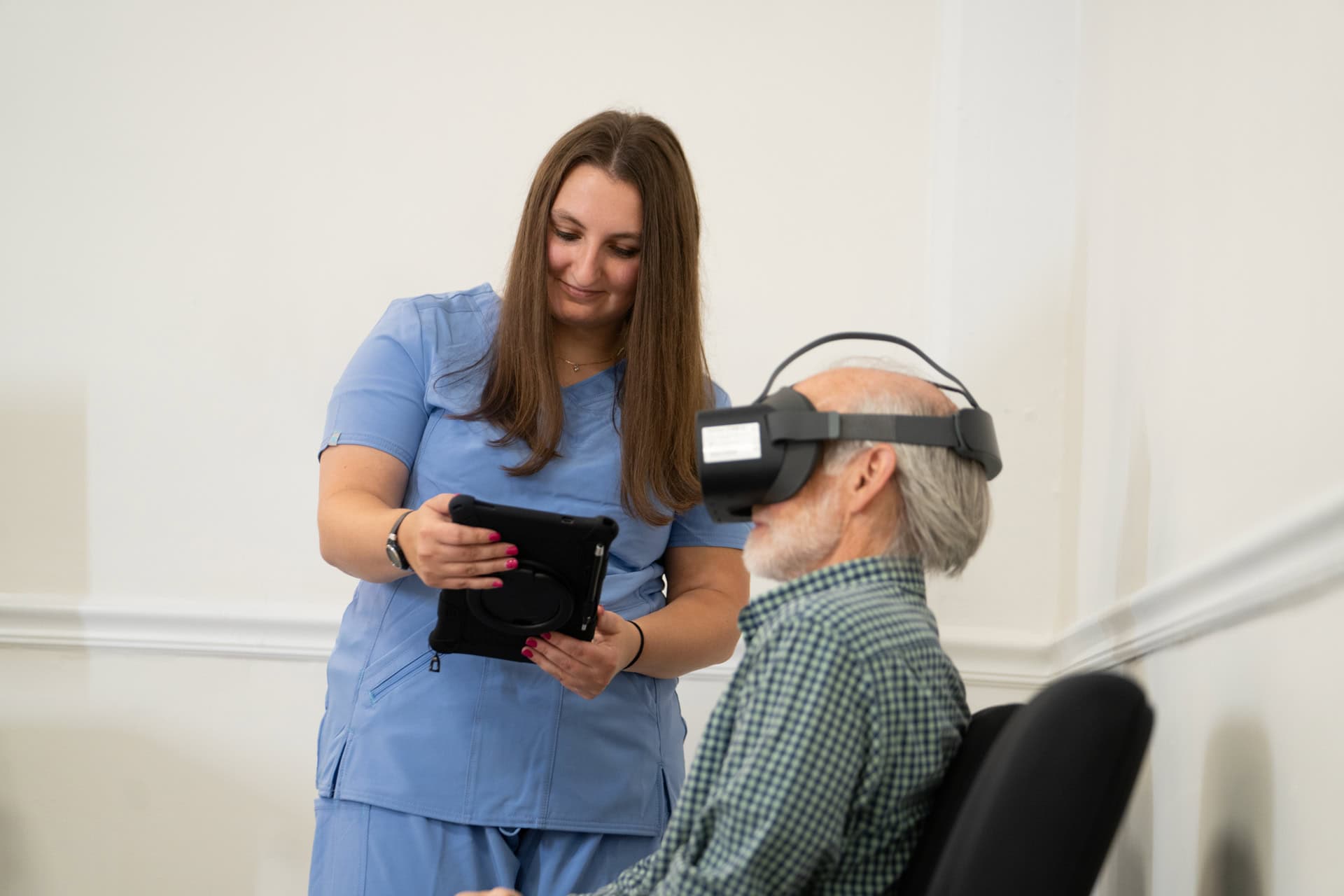
In recent years, virtual reality visual field systems have revolutionized the screening process. Using head-mounted displays, virtual reality tools simulate traditional perimetry environments in a compact, portable format.
VR perimetry and virtual reality perimetry allow clinicians to test each eye individually while tracking fixation, response time, and sensitivity levels. These systems offer several benefits:
-
- Portability – suitable for outreach camps and bedside testing.
- Improved patient comfort – less intimidating than bulky conventional perimeters.
- Enhanced data analytics – AI-driven interpretation and progress tracking.
The integration of virtual visual field assessments provides accurate, repeatable results, ensuring reliable screening even outside the clinic.
Complementary Diagnostic Tools in Modern Eye Care
Accurate diagnosis of ocular diseases depends on combining field testing with other essential investigations. Some critical devices used alongside perimetry include:
-
- Biometry – for measuring axial length, crucial before cataract or refractive surgeries.
- A-Scan (Ascan) – provides ultrasound-based axial length measurement for intraocular lens (IOL) calculation.
- B-Scan (Bscan) – a two-dimensional ultrasound image used to visualize posterior segment structures when media opacities obscure direct view.
- Pachymeter – measures corneal thickness; Pachymetry is vital in glaucoma assessment and refractive surgery planning.
- Keratometer – determines the curvature of the cornea, essential for contact lens fitting and astigmatism evaluation.
- CXL (Corneal Crosslinking) or Corneal Cross-linking – strengthens corneal tissue in conditions like keratoconus by forming new collagen bonds through riboflavin and UV light.
Together, these diagnostic techniques form a complete ocular assessment protocol, where virtual field testing complements anatomical and structural evaluations.
Best Practices for Reliable Confrontation Testing
To ensure accuracy and consistency in confrontation visual field screening:
-
- Maintain consistent lighting – Perform the test in evenly illuminated conditions to avoid false negatives.
- Use equal fixation – Both examiner and patient should maintain steady fixation throughout.
- Test all quadrants – Superior, inferior, nasal, and temporal fields must be assessed individually.
- Compare both eyes – Differences may indicate monocular field defects or neurological issues.
- Document findings carefully – Even if the test is subjective, note any delays or omissions in detection.
When possible, correlate results with VR perimetry or virtual visual field findings for a comprehensive assessment.
Future of Visual Field Testing
With advances in vision, virtual reality, and virtual reality visual field platforms, eye care is shifting toward portable, immersive, and data-driven diagnostics. Combining Confrontation Visual Field Testing with virtual perimetry ensures accessibility in primary care settings while maintaining diagnostic precision comparable to standard automated perimeters.
As integration between virtual reality perimetry and imaging tools like A-Scan, B-Scan, and Pachymetry becomes seamless, clinicians can deliver complete ocular diagnostics efficiently — bridging the gap between traditional methods and modern virtual visual field technology.
Conclusion
The Confrontation Visual Field Test remains a cornerstone of initial vision assessment. However, when enhanced with modern technologies such as VR perimetry and complemented by biometry, pachymetry, and corneal cross-linking procedures, it contributes to a holistic view of visual function and ocular health. The blend of clinical expertise and virtual reality vision testing defines the future of accurate, accessible, and patient-friendly eye care.


